Email: info@forestskinclinic.com
Telephone: 01992 276376
Mobile: 07379 336376
Mobile: 07379 336377
Medical Dermatology
Medical Dermatology
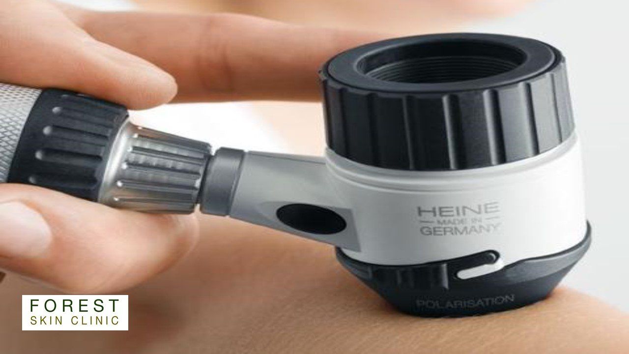
Skin Cancer Screening
Skin Cancer Screening
People with risk factors for melanoma and skin cancer include those who have one or more of the following:
- A history of childhood sunburn
- A fair skin (particularly pale skin and red hair)
- Had high UV exposure in the past
- Lived overseas in a hot climate
- Worked outdoors
- Pursued outdoor hobbies
- A tendency to be a sun-seeker / history of sunbathing
- A history of sunbed use
- Multiple moles
- Multiple atypical moles
- A history of skin cancer
- A history of melanoma
- A family history of melanoma
- A history of immunosuppression
People with more than one of these risk factors may benefit from regular skin cancer screening.
Obagi Medical Skin Care System
Obagi Medical Skin Care System
No matter what your age or skin type, there truly is an Obagi product suitable for each individual.
Obagi products are designed to help minimize the appearance of premature skin ageing, skin damage, hyperpigmentation, acne, and sun damage.
Obagi skincare systems and products are clinically proven to help you revitalise, enhance, and maintain beautiful skin for life.
Obagi System is comprised of a range of topical treatments developed to address a variety of skin concerns by using prescription-strength products to focus on changing the way the skin functions at a cellular level, which speeds up its renewal process, diminishes fine lines and wrinkles, sagging skin, hyperpigmentation, sun damage and acne.
There are several successful systems to transform the skin, including the Obagi Nu-Derm® System, Obagi-C Rx® System, Obagi360™ System, CLENZIderm M.D.™ System, Obagi Professional-C™ System.
You will receive a thorough skin analysis and recommended individualised treatment program that combines professional treatments with home care products to address your skin concerns. Pre-treatment products precondition the skin and accelerate results , whilst post-treatment products minimise complications, soothe the skin and then maintain the results. Professional treatments enhance the results of your home care products.
Email: info@forestskinclinic.com
Tel: 01992 276376
Dermatology Surgery
Dermatology Surgery
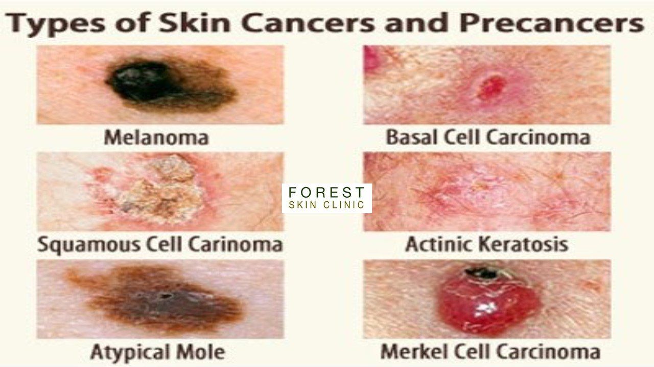
Surgical removal of pre-cancerous and cancerous lesions
Actinic Keratosis
- is at the early stage of the development of a skin cancer - usually treated with Cryosurgery, Photodynamic Therapy or Topical Chemotherapy (a cream), sometimes requires surgical removal via scraping.
Bowen's Disease
- growth of cancerous cell confined to the outer layer of skin - usually treated with Cryosurgery, Photodynamic Therapy or Topical Chemotherapy (a cream) - symptoms can appear similar to other skin conditions such as psoriasis and therefore a biopsy to confirm diagnosis may be required.
Basal Cell Carcinoma
- the most common form of skin cancer accounting for 80% of cases. They rarely spread but need to be treated correctly to prevent them from recurring. Simple excision under local anaesthetic is often a solution. Chemotherapy creams or Photodynamic Therapy are sometimes the more appropriate treatment options.
Squamous Cell Carcinoma
- is the second most prevalent type of skin cancer in the UK. It affects the outer layers of the skin, the epidermis. If left can develop into invasive squamous cell carcinoma where cancer cells grow into the deeper layers of the skin. Simple, wide surgical excision under local anaesthetic is effective. Chemotherapy creams, or photodynamic therapy are not normally effective.
Melanoma
- The most serious type of skin cancer and it is on the increase. This can penetrate all layers of the skin and spread to other organs. In most cases surgical excision is the solution.
Merkel Cell Carcinoma
- is a rare but highly aggressive skin cancer. They tend to invade locally, infiltrating the underlying subcutaneous fat, fascia, and muscle, and typically metastasize early in their natural history, most often to the regional lymph nodes. Surgical excision is the first stage of treatment.
All surgical procedure samples will be sent for analysis and cases discussed at a Multi-dsciplinary meeting to ensure you receive the optimum care.
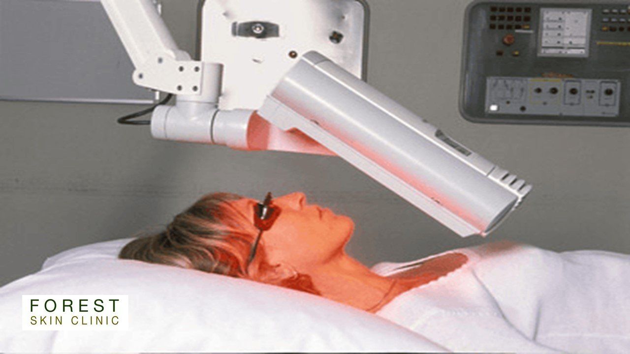
Photodynamic Therapy (PDT)
Photodynamic Therapy (PDT)
For the treatment of Actinic Keratosis, Bowens Disease and Superficial Basal Cell Carcinoma. Also used for skin rejuventation.
Traditional PDT
Each treatment visit comprises of two components, which takes about 3 1/2 hours in total to complete.
Application
– Firstly, the area for treatment is marked and a photosensitising cream applied and a dressing placed to cover the area.
3 hours need to pass to allow time for the cream to be absorbed into the skin.You are free to leave the clinic during this time alternatively refreshments are available if you prefer to stay within the clinic.
Activation
– Once the cream has been absorbed, the dressing is removed and the excess cream cleared from the skin. The treatment area is then placed under the treatment lamp for approximately 10 minutes. This process activates the cream, making it toxic to the cells that have absorbed it, namely the tumour cells. After treatment a simple dressing is applied.
The treatment is repeated the following week for skin conditions that require two treatment sessions.
Some conditions are suitable for Daylight PDT. Your clinician will discuss treatment options with you.
Cryotherapy
Cryotherapy
Removal of pre-cancerous and benign lesions by freezing.
- Actinic Keratosis
- Bowen's Disease
- Superficial Basal Cell Carcinoma
- Seborrhoeic Keratosis
- Viral Warts
- Skin tags
- Verruca
- Early prevention of keloid growth
Treatment is short, perhaps half an hour and recovery equally quick, you can go about your day as normal. One session usually deals with the problem, follow on sessions may be occasionally required. You may be left with a white scar.
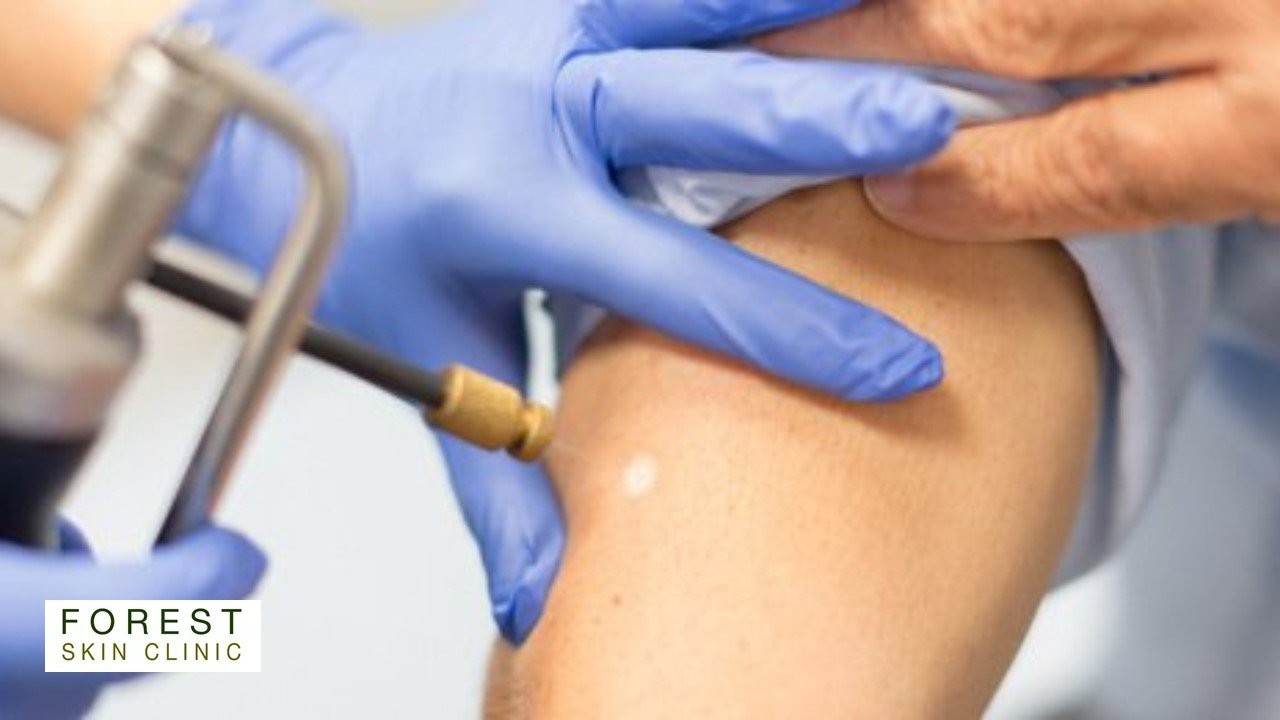
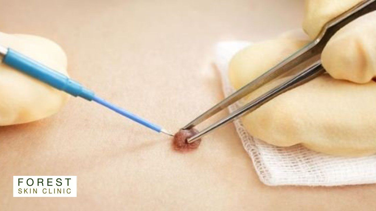
Minor Skin Surgery
Minor Skin Surgery
For the removal of benign lesions
- Large Seborrhoeic Keratosis
- Multiple Skin tags
- Cherry angiomas - benign lesions caused by enlarged capillaries
- Benign protruding moles, catching on clothes
All lesions will be sent for analysis to reassure you of their benign nature.
These procedures will leave a scar.
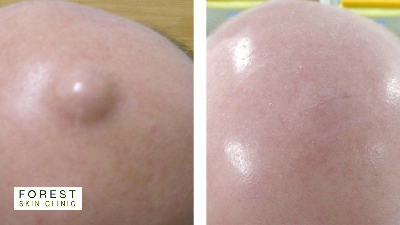
Cyst Removal
Cyst Removal
A skin cyst is a round, dome-shaped lump. It's yellow or white, often with a small dark plug through which you might be able to squeeze out pus.
Cysts can range in size from smaller than a pea to a few centimetres across. They grow slowly.
Skin cysts do not usually hurt, but can become tender, sore and red if they become infected.
Foul-smelling pus coming out of the cyst is another sign of infection.
Epidermoid cysts (one of the main types) are commonly found on the face, neck, chest, shoulders or skin around the genitals.They affect young and middle-aged adults, and are particularly common in people with acne. They do not usually run in families.
Pilar cysts form around hair follicles. They're often found on the scalp. Pilar cysts typically affect middle-aged adults, particularly women. Unlike epidermoid cysts, they run in families
Do not be tempted to burst the cyst. If it's infected, you risk spreading the infection, and it can grow back if the sac is left underneath the skin.
During a cyst removal, a local anaesthetic is used to numb the skin. A tiny cut is then made in the skin and the cyst is squeezed out. The sac is then removed to prevent the cyst from growing back.
This procedure will leave a scar.
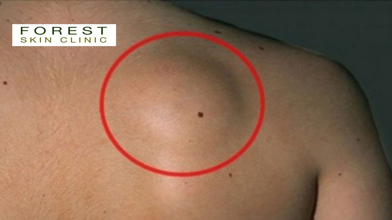
Lipoma Removal
Lipoma Removal
Lipomas are soft, fatty lumps that grow under your skin.
They're harmless and don't usually need any treatment.
Lipomas are benign and are caused by an overgrowth of fat cells. They can grow anywhere in the body where there are fat cells.
Removing a lipoma under these circumstances is regarded as cosmetic surgery
Lipomas are common. They:
- feel soft and squishy
- can be anything from the size of a pea to a few centimetres across
- may move slightly under your skin if you press them
- aren't usually painful
- often appear on your shoulders, chest, arms, back, bottom or thighs
- grow slowly
Procedures used to remove lipomas will depend on the nature of the lipomas and its location on your body.
During a lipoma removal, a local anaesthetic is used to numb the skin. A tiny cut is then made in the skin and the lipoma and the connecting fibres are squeezed out.
This procedure will leave a scar.
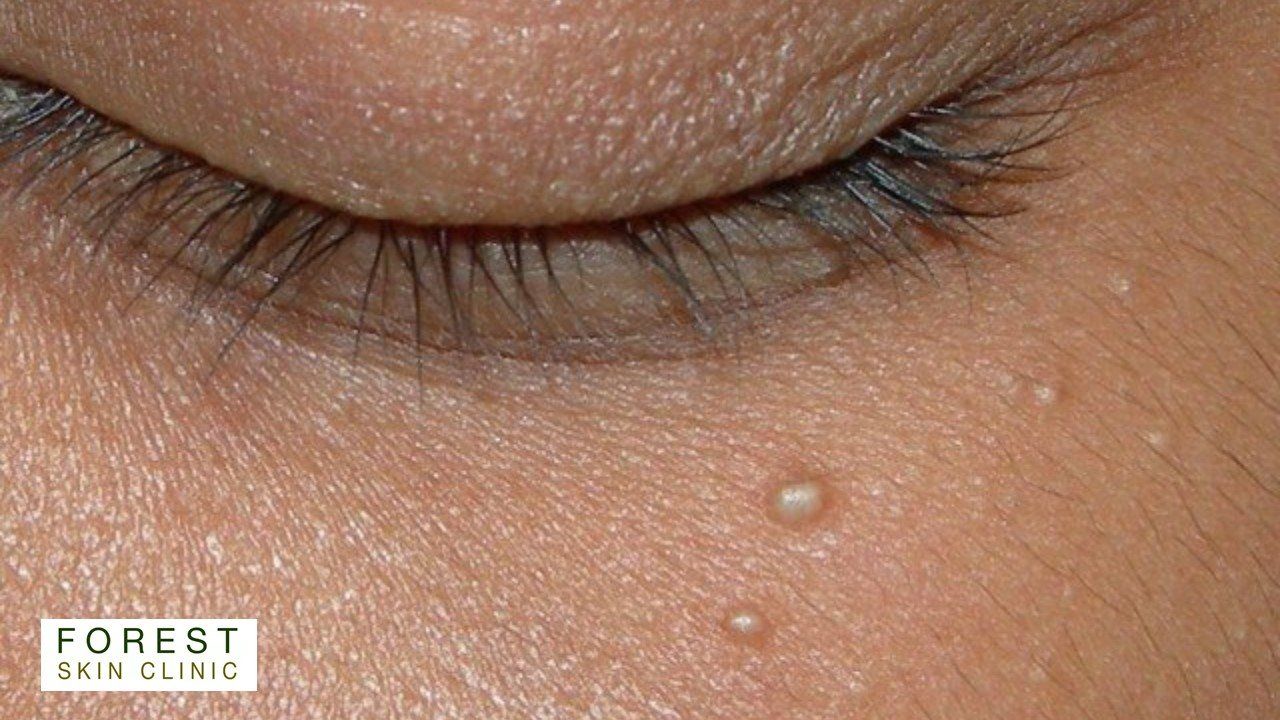
Milia Removal
Milia Removal
Milia are small, white, yellowish, or skin coloured spots, which form when keratin, the protein which makes hair and our outer skin, clog up the sweat ducts, whilst secondary milia can occur after trauma, or using the wrong skin cream.
They feel firm and rarely look inflamed.
There are also less common types. Multiple eruptive milia brings clusters of spots, milia en plaque can see the skin inflamed, or raised. Ensuring correct diagnosis is the key to treatment
Although they are harmless, many people with milia would prefer to remove them.
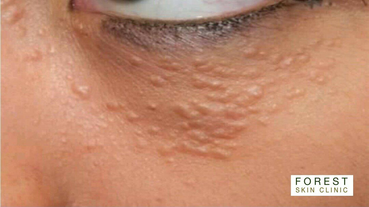
Syringoma Removal
Syringoma Removal
A syringoma is a benign, or non-cancerous, growth caused by overactive sweat glands. Syringomas usually develop on the neck, upper cheeks, and the lower region of the eyes, but occasionally they grow on the abdomen, armpit, scalp, bellybutton, and genitals.
In most cases, syringomas are harmless and do not cause symptoms. Rarely, however, some individuals with syringomas experience extreme pain and itchiness, especially when sweating.
Syringomas mostly develop in early adulthood, between the ages of 25 and 30.
Syringomas are linked to several different medical conditions, including diabetes.
Though rare, some people have a genetic predisposition towards developing them.
Once syringomas have been diagnosed, there is usually no reason to treat them.
Syringomas are small papules, or firm bumps, that are about 1 to 3 millimeters wide. The papules usually grow in small groups and are typically:
- yellow
- brown
- pale pink
- skin-toned
Syringoma clusters tend to be symmetrical, meaning the same pattern appears on both sides of the body in the same place.
Syringomas develop when sweat duct cells in the outermost layer of skin overgrow or sweat glands overreact, forming tumors or abnormal tissue growths. The sweat ducts are tubular structures that carry perspiration from the sweat gland to the skin's surface for release and cooling.
Electrosurgery
During electrosurgery, electrical currents are concentrated and sent through the syringoma. These currents destroy abnormal tissues and damage blood vessels.
Dermabrasion
Dermabrasion involves physically remove and even out the top layers of the skin. This procedure is often unsuitable for syringomas that are rooted deep in a person's skin.
According to the American Society for Dermatologic Surgery, a 50 percent improvement in the targeted skin condition is considered a successful dermabrasion surgery.
Chemical peels
Certain chemicals, most commonly trichloroacetic acid, can be applied directly to the syringoma, usually causing it to dry up and fall off. When done by a trained nurse or doctor chemical peels often do not cause scarring.
Surgical excision
In some cases, syringomas need to be surgically removed using traditional cutting, scraping, and peeling instruments. Surgery is often the last resort option, given excision almost inevitably leads to scarring and tissue damage.
Excision may be the only option for papules that are embedded deep within a person's skin. Surgeons will use sutures or stitches to close the resulting open wound.
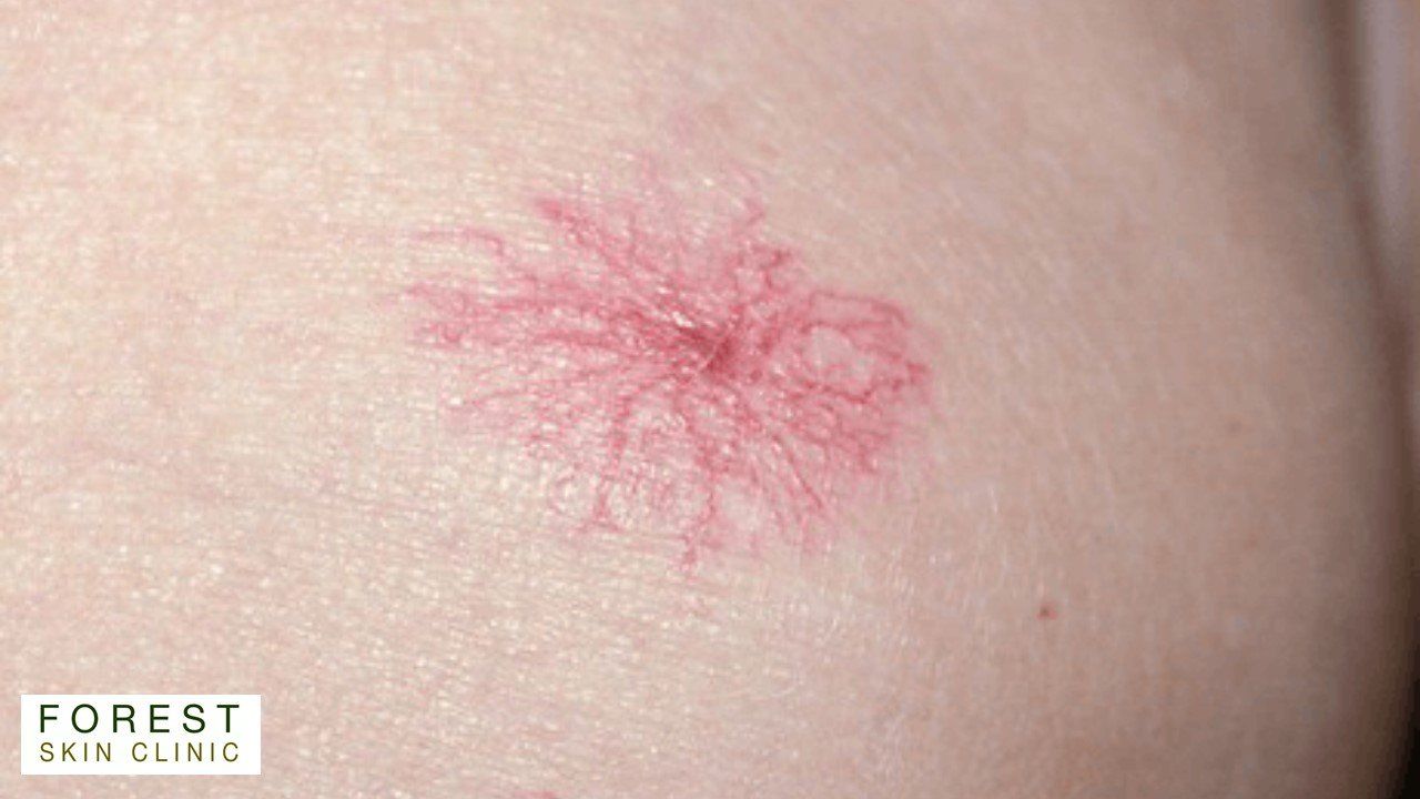
Spider Naevus Removal
Spider Naevus Removal
A spider naevus is a collection of small, dilated arterioles (blood vessels) clustered very close to the surface of the skin. The cluster of vessels is web-like, with a central spot and radiating vessels.The thin vessels form a web-like shape and are red, blue, or purple in color. When you apply pressure, they will disappear and then reappear because blood is flowing back into the vessels.
Spider naevus commonly occurs when you have a lot of oestrogen in your system, as is the case with chronic liver disease or during pregnancy as well as taking hormonal contraceptives.. The older you are, the more likely you are to get spider nevi as the valves in your blood vessels to weaken.
Being in the sun, especially if you are fair-skinned, can cause spider nevi to form on your face.
Hot point cautery is used to coagulate the lazy blood vessel at the centre that is causing the spider naevus. You may require a couple of treatments.
Microsclerotherapy
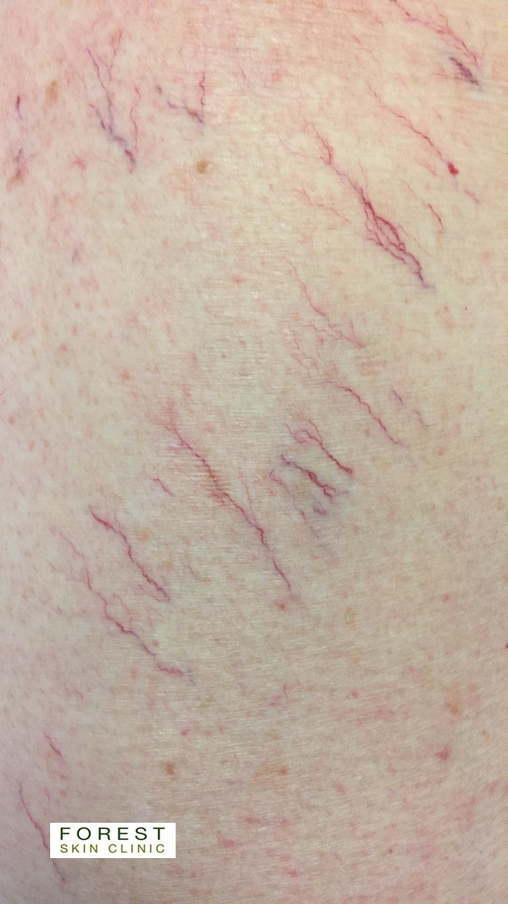
Microsclerotherapy
Thread veins are unsightly very small, abnormally dilated blood vessels. They are red/blue in colour and lie very close to the surface of the skin. Usually they cause no physical problems, occasionally they may cause discomfort and itching.
Microsclerotherapy is most effective in the treatment of thread veins on the legs, and involves the injection of a solution, or ‘sclerosant’, with a very fine needle into the problem blood vessel. The injected solution is designed to destroy the endothelium triggering thrombosis (clotting) and subsequent fibrosis (hardening), causing the vessel to close and fade away. The use of graduated compression hosiery supports and promotes the healing process, minimising undesirable side effects such as the development of post-sclerotherapy thrombi (clots) and general inflammation. A ten minute walk immediately after treatment, and then 72 hours continuous compression. This is followed by compression for two weeks during the day.
During your initial consultation you will be assessed in a standing position, this enables the practitioner to fully assess your legs. This can be done by looking and feeling the legs. A handheld Doppler machine is used to listen to the blood in your veins . This will check the flow of blood in the veins and check for any problems both superficially and deeper in your legs.
The practitioner should at this time be able to tell you how many sessions you will need and their cost. This varies according to the quantity and type of veins requiring treatment. Most sessions take between ½ to 1 hour. You should budget for at least two treatments.
The results after one treatment vary considerably from one client to another. Some veins may disappear completely whilst others may only fade and some remain unchanged. On completion of a successful treatment you can expect an improvement in the overall appearance of 60 – 80%. We hope that you will see enough improvement to increase your confidence in the way your legs look.
Your susceptibility to thread veins does mean that other thread veins may emerge over a period of time. In most cases, further treatments will be necessary. Some clients prefer to return each year whilst others may come back when more veins appear after a few years.
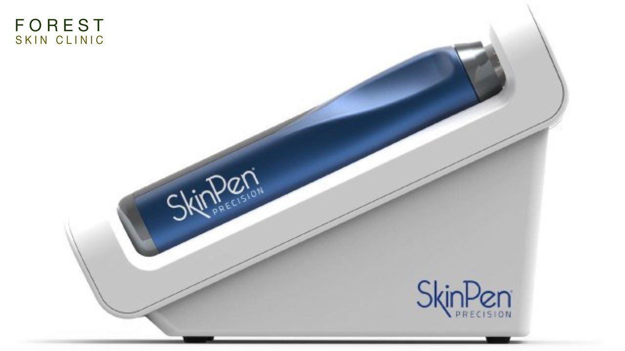
SkinPen Precision Microneedling for the treatment of Ageing, Wrinkles, Scars
SkinPen Precision Microneedling
for the treatment of Ageing, Wrinkles, Scars
The SkinPen Precision Procedure:
- A comfortable experience
- A short procedure time, approximately 40 minutes
- A plan for optimal results, typically 3-6 procedures
- Mild post-procedure effects, similar to a mild to moderate sunburn
- Effective on all body parts including face, neck and décolletage
- Safe for all skin types, light to dark
- Quick 48 hour recovery time. Little to no downtime
- Beautiful, lasting results
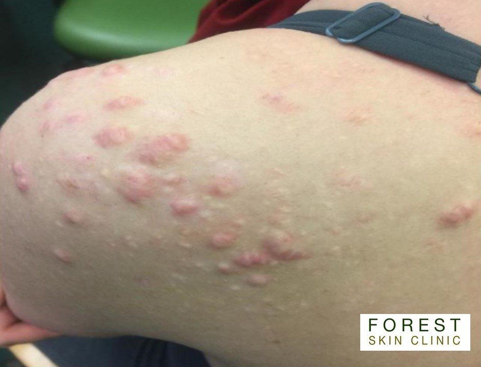
Keloid Scar Treatment
Keloid Scar Treatment
A keloid scar is an enlarged, raised scar that can be pink, red, skin-coloured or darker than the surrounding skin.
They can develop after very minor skin damage, such as an acne spot or a piercing, and spread beyond the original area of skin damage.
Keloid scars are more common on the upper chest, shoulders, head (especially the earlobes after a piercing) and neck, but they can happen anywhere.
Anyone can get a keloid scar, but they're more common in people with dark skin, such as people from Africa and African-Caribbean and south Indian communities.
Experts do not fully understand what causes keloid scars, but they happen when there's overproduction of collagen (the skin's protein).
They're not contagious or cancerous.
If you have had a keloid scar before, you're more likely to get another.
There are several treatments available, but none have been shown to be more effective than others.
Treatment can be difficult and is not always successful.
Treatments that may help flatten a keloid scar include:
steroid injections
applying steroid-impregnated tape for 12 hours a day
applying silicone gel sheeting for several months
freezing early keloid scars with liquid nitrogen to stop them growing.
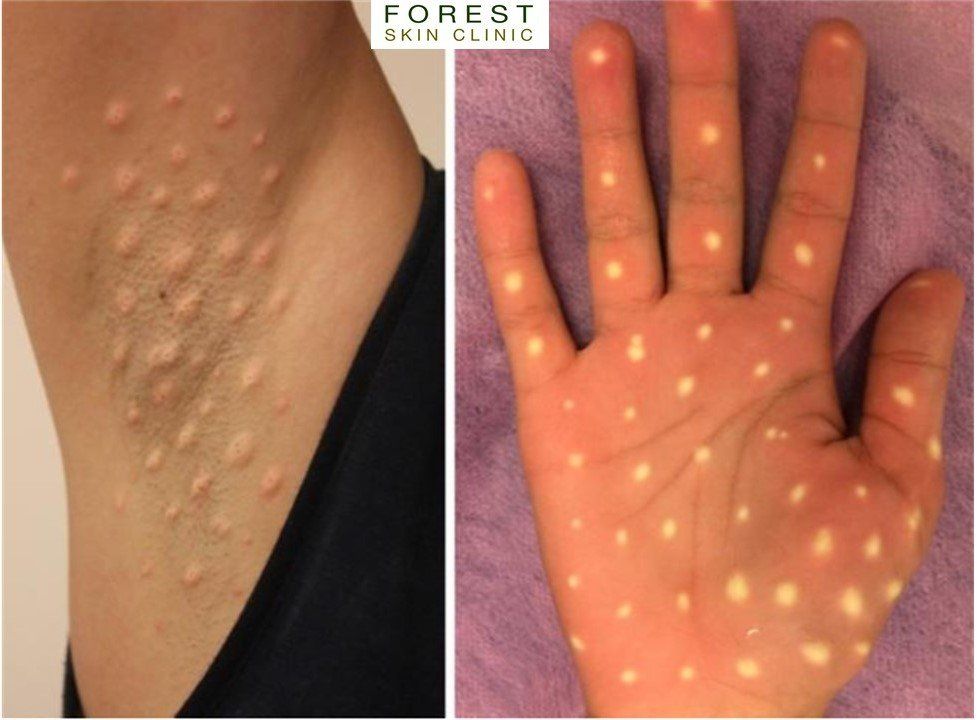
Hyperhidrosis - excessive sweating
Hyperhidrosis - excessive sweating
Botulinum toxin is delivered by multiple intradermal injections to the affected areas.
It acts by inhibiting acetylcholine release from the sympathetic cholinergic nerve terminals that innervate sweat glands.
Botulinum toxin is licensed for the treatment of axillary hyperhidrosis and may also be used for palmar, plantar, and craniofacial hyperhidrosis (treatment is more painful in these areas). The effect may last for 6–9 months.
Adverse effects include pain during injections and compensatory sweating. Transient muscle weakness and loss of fine motor control have also been reported.

Company Number 12054118
Registered Address:
219 High Road
North Weald
CM16 6ED